Anglický jazyk
8.3 Medical English - The respiratory system
The respiratory system describes how breathing = respiration and gas exchange take place in the human body.
Air enters the body through the nose and passes through the nasal cavities which are lined with mucous membrane and fine hairs called cilia. These hairs help filter out foreign bodies and warm and moisten the incoming air. Behind the nasal cavities there are the paranasal sinuses which are located within the cranium and filled with air. They also have mucous membrane lining and their function is to provide the lubricating fluid mucus as well as to lighten the skull bones and help produce sound.
Then the air reaches the throat = pharynx. We can divide it in three parts top-down: the nasopharynx, which contains the adenoids. They might obstruct airways if enlarged. Closer to the mouth is the oropharynx where the tonsils are located. And the third part is the hypopharynx = laryngopharynx. Here the pharynx divides in two passageways: the larynx for air, and the oesophagus for the food. The larynx contains the vocal cords and cartilages for support. The vocal cords are the key organ that lets people produce sounds. Those are made as air is expelled from the lungs and the cords vibrate. Their tension determines the high or low pitch of the voice.
Epiglottis is the little valve that prevents food from entering the airways. It is a flap of cartilage attached to the root of the tongue which acts as a lid over the larynx. It lies over the mouth of the larynx which is called glottis. When we swallow the glottis moves under the epiglottis automatically and closes off the larynx so that food cannot enter it.
After passing the larynx the air goes on to the trachea = windpipe. In the region of mediastinum, the trachea divides in two branches called bronchi. Each bronchus leads to a separate lung and subdivides into smaller and finer tubes. The tiniest tubes are called bronchioles. At their end there are clusters of air sacks called alveoli. They have very thin walls which allow for the exchange of gases between the alveolus and the capillaries. The blood which flows through the capillaries accepts oxygen from the alveolus and deposits carbon dioxide in the alveolus to be exhaled.
Each lung is enveloped in a double folded membrane called the pleura. Its outer layer is the parietal pleura and the inner layer is the visceral pleura. The two lungs are not identical. The right lung is divided into three lobes while the left one in only two lobes. The lungs extend from the collar bone to the diaphragm in the thoracic cavity. The diaphragm is a muscular partition which separates the thoracic and abdominal cavities. The diaphragm contracts and descends with each inspiration = inhaling, breathing in, and relaxes and ascends with expiration = exhaling.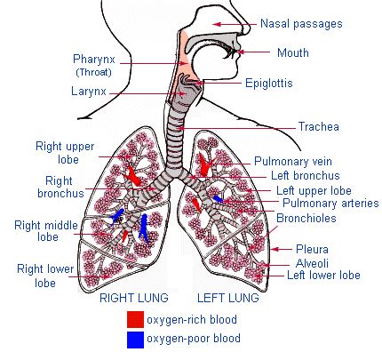
|
gas exchange
|
výměna plynů
|
|
enter
|
vstoupit do
|
|
nasal cavities
|
nosní dutiny
|
|
cavity, sinus
|
dutina
|
|
foreign bodies
|
cizí tělesa
|
|
warm and moisten
|
ohřát a zvlhčit
|
|
lighten
|
odlehčit
|
|
adenoids
|
žlázy
|
|
airways
|
dýchací cesty
|
|
obstruct
|
blokovat
|
|
enlarged
|
zvětšený
|
|
tonsils
|
mandle
|
|
vocal cords
|
hlasivky
|
|
expell
|
vyloučit, vytlačit
|
|
tension
|
napětí
|
|
pitch
|
tón
|
|
gadget
|
pomůcka
|
|
flap
|
cíp
|
|
lid
|
poklička
|
|
windpipe
|
průdušnice
|
|
tiny
|
drobný
|
|
cluster
|
hrozen, trs
|
|
alveolus
|
plicní sklípek
|
|
deposit
|
odložit
|
|
carbon dioxide
|
oxid uhličitý, CO2
|
|
envelope
|
obalit
|
|
lobe
|
lalok
|
|
diaphragm
|
bránice
|
|
partition
|
přepážka
|
|
ascend – descend
|
stoupat – klesat
|
|
inspiration – expiration
|
nádech – výdech
|
Some complications connected with the respiratory system
A cough is a common symptom of upper respiratory tract infection and lung disease. A cough may be productive = loose, where the patient coughs up sputum= phlegm, or non-productive = dry, where there is no sputum. Sputum may be clear or white (mucoid), yellow due to the presence of pus (purulent), or blood-stained (as in haemoptysis). Lung diseases are often connected with dyspnoea, which is basically getting short of breath.
Doctors examine the patient's breathing by auscultation = listening to the chest with a stethoscope. There are two main added sounds: crackles which sound like hairs being rubbed together and suggest the presence of fluids in the lungs, and wheezes which are more musical, perhaps like whistling, and indicate narrowing of the airways. This is typical for asthma. The sound heard when the pleural surfaces are inflamed as in pleurisy is called a pleural rub. The doctor might ask the patient to say some words to check their vocal resonance. It may be increased as in pneumonia or decreased as in pneumothorax.
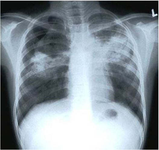
There are more lung diseases such as tuberculosis which is quite infectious and life threatening although patients might live many years after contracting it.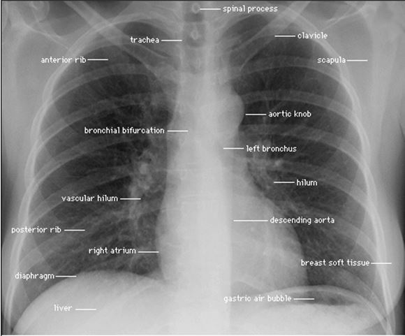
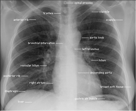
Healthy lungs.
Source: http://4.bp.blogspot.com/-9JadtjswWKY/UBqypjyWhiI/AAAAAAAAABs/eaysS4wmUjY/s1600/X-ray-Chest.gif
Lungs that suffer from tuberculosis.
|
cough
|
kašel, kašlat
|
|
phlegm [flem]
|
hlen
|
|
pus
|
hnis
|
|
auscultation
|
poslech
|
|
whistle
|
pískat
|
|
narrow
|
úzký, zúžit
|
|
inflamed
|
zanícený
|
|
get short of breath
|
nemoci popadnout dech
|
|
threaten
|
hrozit, ohrožovat
|
|
contract (a disease)
|
onemocnět čím
|